Conference Agenda
Two days of cutting-edge presentations, live demonstrations, and interactive discussions.
Free paper presentation session also available at 7:00 - 7:30 on both days.
THURSDAY, DECEMBER 11, 2025
06:45-
30 minutes
REGISTRATION
07:30 - 08:00
30 minutes
FROM TMR TO RPNI
Moderator: Chih-Sheng La (賴志昇) / Tommy, Nai-Jen Chang (張乃仁)
08:00 - 08:30
30 minutes
PRESIDENTIAL INVITED LECTURE
Moderator: Cheng-Hung, Lin (林承弘)
08:40 - 09:00
20 minutes
PREOP DISCUSSION
09:00 - 11:00
120 minutes
LIVE SURGERY
LIVE SURGERY (BROADCAST TILL 14:00)
Demonstrated by Oskar Aszmann
Assisted by Harvey Chim
Witness cutting-edge surgical techniques in real-time
Yu-Te Lin (林有德) / Chung-Chen Hsu (許聰政) / Yu-Huan Hsueh (薛宇桓) / Changsik John Pak / Jason Shih Hoellwarth

Oskar Aszmann
Department of Plastic and Reconstructive Surgery, Medical University of Vienna, Austria
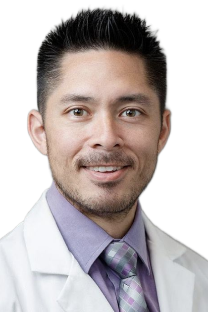
Harvey Chim
Department of Plastic and Reconstructive Surgery, University of Florida, USA
11:00 - 12:00
60 minutes
TSRM/JSRM/KSM JOINT SESSION I
Moderator: Chun-Ta Li (李俊達) / Chih-Hung Lin (林志鴻)/ Johnson, Chia-Shen Yang(楊家森)
12:10 - 13:00
50 minutes
會員大會/LUNCH
13:00 - 14:00
60 minutes
KEYNOTE
BIONIC LIMB VS HAND TRANSPLANTATION
Moderator: Hao-Chih Tai (戴浩志)/ Shih-Heng Chen (陳思恒)
Panel discussion: Transplantation vs Bionic reconstruction
Paul Cederna
Section of Plastic and Reconstructive Surgery, Department of Surgery, University of Michigan, USA
/profilePic.png)
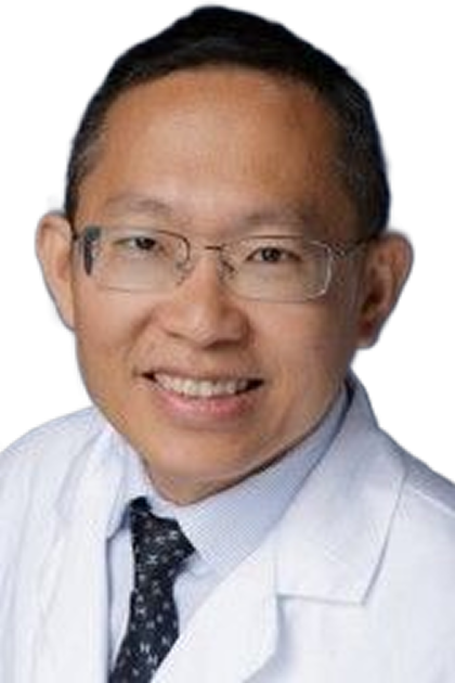
Cheng-Hung Lin (林承弘)
(林承弘)Department of Plastic and Reconstructive Surgery, Chang Gung Memorial Hospital, Taiwan
14:00 - 15:10
70 minutes
CME
NEUROMA AND PHANTOM PAIN MANAGEMENT (CME)
Moderator: Chih-Hao Chang (張志豪) / Honda Hsu (許宏達) / Wen-Chih Liu (劉文智)
15:20 - 15:30
1.0 minutes
COFFEE BREAK
15:30 - 17:30
120 minutes
HANDS-ON
HANDS-ON ULTRASOUND NEUROGRAPHY
Moderator: Hsin-I Tsai (蔡欣怡) / Shih-Jyun Shen (沈士鈞) / Yen-Po Lin (林彥伯) / Shao-Chih Hsu (許韶芝) / Liang-Jun Ou- Yang (歐陽良俊)
FRIDAY, DECEMBER 12, 2025
07:00 - 07:30
30 minutes
BREAKFAST AND EXHIBITS
07:30 - 08:30
60 minutes
MAJOR LIMB TRAUMA RECONSTRUCTION & BEYOND?
Moderator: Ying-Sheng Lin (林穎聖) / Suan-Chuan Chao (趙崧筌)
08:30 - 09:00
30 minutes
PREOP DISCUSSION
09:00 - 11:00
120 minutes
LIVE SURGERY
LIVE SURGERY (BROADCAST TILL 14:00)
Demonstrated by Paul Cederna
Assisted by Theodore Alexander Kung
RPNI demonstration: transhumeral amputation
Treatment of painful neuroma
Treatment of phantom pain
Moderator: Honda Hsu (許宏達) / Yun-Jui Lu (呂昀叡) / Wen-Chih Liu (劉文智) / Changsik John Pak / Jason Shih Hoellwarth
Paul Cederna
Section of Plastic and Reconstructive Surgery, Department of Surgery, University of Michigan, USA
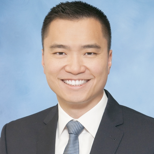
Theodore Alexander Kung
Plastic Surgery, Department of Surgery, University of Michigan, USA
11:10 - 12:00
50 minutes
TSRM/JSRM/KSM JOINT SESSION II
Moderator: Hung-Chi Chen (陳宏基) / Chih-Hung Lin (林志鴻) / Pao-Jen Kuo (郭寶仁)
12:10 - 13:00
50 minutes
LUNCH SEMINAR
3M LUNCH SEMINAR
13:00 - 14:00
60 minutes
KEYNOTE
BIONIC LIMB AND OSSEOINTEGRATION
Moderator: Jung-Hsien Hsieh (謝榮賢) / Cheng-Hung Lin (林承弘) / Yu-Huan Hsueh (薛宇桓)
Oskar Aszmann
Department of Plastic and Reconstructive Surgery, Medical University of Vienna, Austria
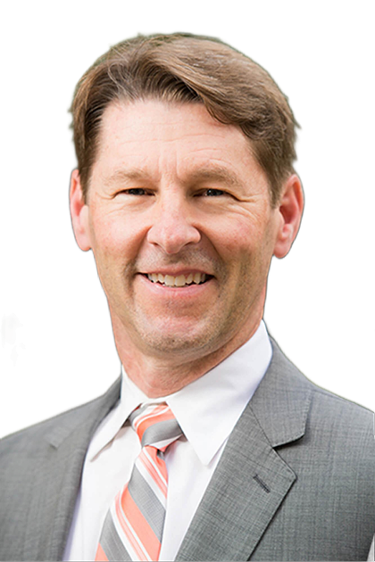
Jason Shih Hoellwarth
Department of Orthopedic Surgery, Hospital for Special Surgery, USA
14:00 - 15:10
70 minutes
ADVANCEMENT IN BIONIC LIMBS/EXOSKELETION AND BIOTECHNOLOGY
Moderator: Yur-Ren Kuo (郭耀仁) / Chieh Huei Huang (黃傑慧)
VR rehab program for upper and lower amputees
15:10
CLOSING SESSIONS
Awards
Wrap-up